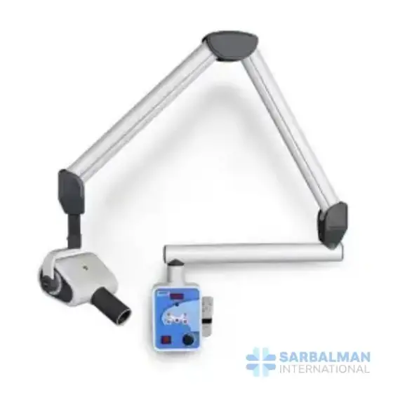
RVG Unit
Free!
An RVG unit is a digital intraoral X-ray system that delivers sharp images to your computer in seconds. It provides adjustable exposure, a sensitive CMOS sensor, and software tools for measuring and sharing. Ideal for bitewings, periapicals, and endodontic or implant checks, it speeds diagnosis, reduces consumables, and supports lower radiation compared with film. Choose it to modernize your workflow and improve chairside decision-making.
Description
An RVG unit (radiovisiography unit) is a digital intraoral X-ray system that captures sharp dental images directly on a computer in seconds. The setup typically includes a wall-mounted X-ray generator with a positionable arm, a high-sensitivity CMOS sensor, and a control panel or foot switch. Because the sensor is more sensitive than film, useful images can be achieved with lower exposure, while software tools allow instant viewing, enhancement, measurement, and sharing across the clinic.
Key features and benefits
• Instant image capture with real-time preview for faster diagnosis and chairside discussion
• High-contrast, low-noise images suitable for caries detection, endodontic measurements, periodontal assessment, and implant planning
• Adjustable exposure parameters (kVp, mA, time) with adult/child presets to match tooth region and minimize dose
• Ergonomic, rounded-edge sensors in common sizes; sealed housing used with disposable barriers for infection control
• Long, counterbalanced arm for accurate tube head positioning and repeatable projections
• Image tools: zoom, filters, annotations, and measurement calibration; export in common formats and DICOM compatibility for most imaging/PMS software
• Digital workflow eliminates chemicals, film, and scanners, reducing costs and environmental impact
• Durable construction and simple maintenance compared to film processors and plate scanners
Typical applications
• Bitewing images for proximal caries and crestal bone levels
• Periapicals for root morphology, periapical pathology, and endodontic working length
• Occlusal views and check shots during restorative or surgical procedures
How it compares
• Versus film: faster, consistent results, lower operating costs, and generally lower radiation for equivalent diagnostic quality.
• Versus phosphor plate systems: no scanning step, fewer moving parts, and immediate images; plates remain useful for larger fields.
Quality and compliance notes
• Select units that document electrical and radiation safety to recognized standards (for example, IEC 60601 series) and meet local radiation regulations.
• Use rectangular collimation and technique guides for best image quality and dose management.


