Ultrasound Machine
Free!
An ultrasound machine is a diagnostic imaging system that uses high-frequency sound waves to create real-time pictures of organs, tissues, and blood flow. It’s widely used for pregnancy scans, abdominal and cardiac exams, vascular checks, and bedside emergency assessment. With no ionizing radiation, it’s ideal for safe, repeatable monitoring and quick clinical decisions in hospitals and clinics.
Description
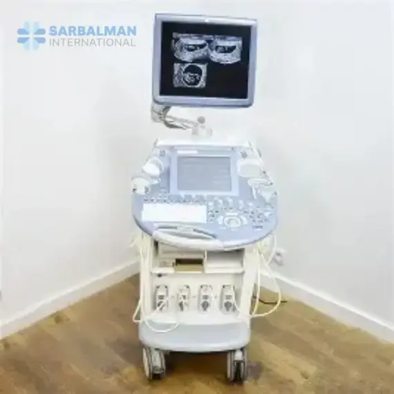
An Ultrasound Machine is a medical imaging device that sends high-frequency sound waves through a handheld probe (transducer) and converts the returning echoes into live images on a monitor. Because the scan happens in real time and does not use ionizing radiation, ultrasound is a trusted first-line tool for safely viewing soft tissues, organs, and fluid-filled structures in patients of all ages.
Key features and benefits include:
-
Multiple imaging modes: 2D/B-mode for anatomy, M-mode for motion (especially cardiac), and Doppler/Color Doppler for assessing blood flow and vessel or heart performance.
-
Clear soft-tissue visualization: Helps clinicians detect abnormalities quickly without exposing patients to radiation.
-
Interchangeable probe options: Different transducers support abdominal, obstetric/gynecologic, cardiac, vascular, and musculoskeletal scanning.
-
Point-of-care portability: Many systems are designed for bedside use, enabling faster decision-making in emergency and critical care settings.
Ultrasound machines are used in hospitals, diagnostic centers, maternity clinics, emergency rooms, and ICUs. Typical applications include fetal monitoring, liver and kidney evaluation, thyroid and breast checks, echocardiography, vascular studies, musculoskeletal imaging, and image-guided procedures such as biopsies or fluid drainage.
Compared with X-ray or CT, ultrasound is safer for repeat monitoring and delivers immediate bedside results. Compared with MRI, it’s faster and more accessible, though less effective for imaging through bone or air-filled areas like lungs.